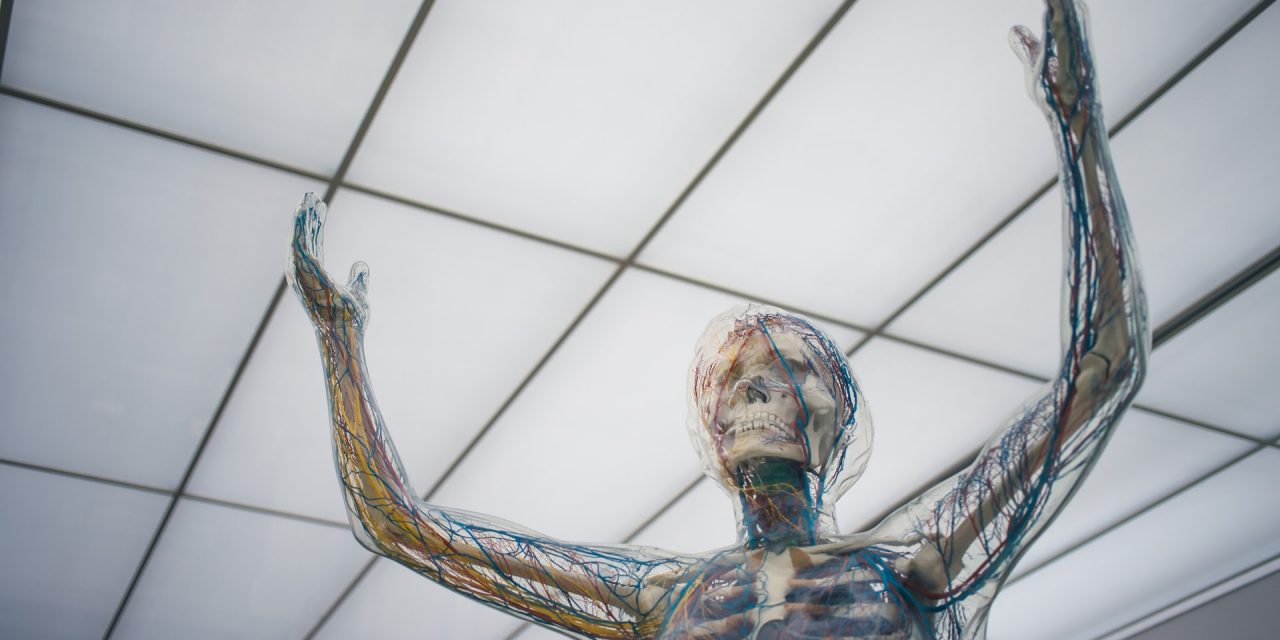
With engineering and medicine combined, ultrasound technologies help physicians and medical experts see more precise and accurate pictures of the internal parts of the human body.
2D PROBE
2D probe repair is the most common one probe used. It uses high-frequency soundwaves for the probes to scan the inside of the body and produce images.
The probe repair requires meticulous engineering skills. France-based repair company, Probe Repair Services (PRS), can conduct repairs on faulty and broken 2D probes. The repair types include membrane replacement, strain relief replacement, wiring sheath replacement, probe wiring replacement, and many more.
The PRS provides repair services for various brands of 2D ultrasound probes, including GE, Siemens, Philips, Toshiba, Hitachi Aloka, to name a few.
PRS has various test machines to determine mechanical issues of the probes, which are typically not detected by electronic testers.
A warranty of three to six months is given, depending on the type of probe.
A GE Ultrasound Repair and Service offers repair of equipment with cutting-edge technology. Their engineers cater to GE ultrasound equipment such as GE Vivid I ultrasound machine and GE Logiq E ultrasound unit, to name a few.
3D AND 4D PROBE
Similar to regular ultrasounds, the 3D and 4D ultrasound probes rely on soundwaves to detect the baby inside the womb of a pregnant woman. Obstetrician-gynecologists, commonly known as OB-GYNE, usually use these probes for ultrasounds.
The 3D probes produce a three-dimensional image, whereas the 4D ultrasound probes create a more realistic live video effect.
The 3D and 4D probes are also known as abdominal ultrasound or sonograms. The probe is placed against the abdominal area to scan the image of the inside of the womb.
PRS can conduct repairs such as dome replacement, motor repair, oil and sealing, and electronic connector repair to limited brands, including GE, Siemens, Philips, and Toshiba.
TRANSESOPHAGEAL PROBE
Transesophageal echocardiography (TEE) is an alternative process of conducting ultrasound on the heart. In TEE, the probe has a specialized ultrasound transducer at the tip which is inserted into the esophagus.
The TEE probe can produce images of the heart and chest area. Its advantages are that it produces clearer images of the heart area that enables experts to identify if there are complications present.
It is usually hard to produce images of the heart because of the chest wall. The TEE probe overcomes this problem. Since the heart is placed just a millimeter away from the esophagus, it is much easier to produce clearer images when the tip of the probe is inserted.
There are many instances when a TEE probe requires repair. Patients who undergo this type of ultrasound bite on the TEE probe, which can lead to deterioration.
Poor care and maintenance can also reduce the quality of the TEE probes.
The PRS provides expert repair of TEE probes. PRS professional technicians are equipped in replacing parts of the probe, such as the head of the TEE transducer.
Aside from that, PRS can also provide loan ultrasound equipment during the period of repair.


